Lauge-Hansen Klassifikation
Beste Darstellung auf radiologyassistant.nl 🌟

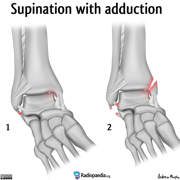
Supination-Adduktion
- stage 1: without medial malleolar fracture
- stage 2: with oblique or vertical medial malleolar fracture
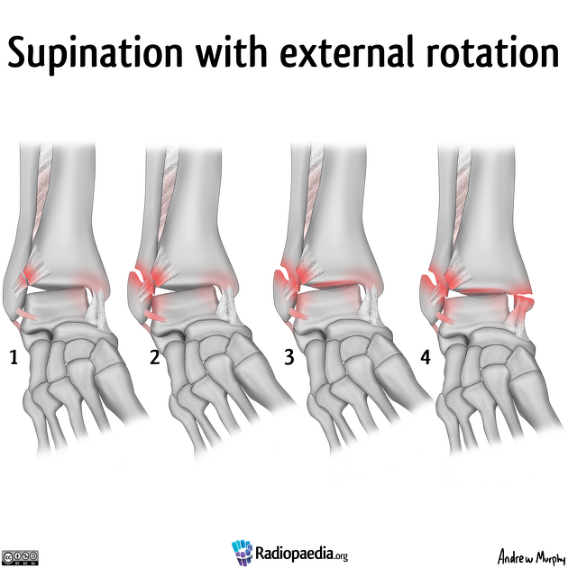
Supination-Eversion (40-70%)
- stage 1: the anteroinferior tibiofibular ligament is torn or avulsed
- stage 2: the talus displaces and fractures the fibula in an oblique or spiral fracture, starting at the joint
- stage 3: tear of the posteroinferior tibiofibular ligament or fracture posterior malleolus
- stage 4: tear of the deltoid ligament or transverse avulsion fracture medial malleolus
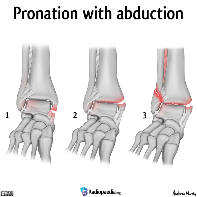
Pronation-Abduktion
- stage 1: deltoid ligament disruption or transverse medial malleolus fracture
- stage 2: posterior malleolus fracture
- stage 3: oblique fibular fracture above the level of the joint, in a low medial high lateral fracture plane
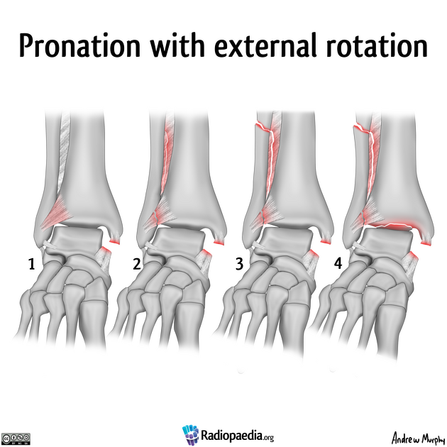
Pronation-Außenrotation
- stage 1: deltoid ligament rupture, which may appear occult or as medial mortise widening, or transverse avulsion fracture of the medial malleolus
- stage 2: involvement of the AITFL with extension into the interosseous membrane results in widening of the distal tibiofibular distance
- stage 3: a spiral or oblique fibular fracture (>6 cm) above the talotibial joint
- stage 4: involvement of the posterior inferior tibiofibular ligament (PITFL), or posterior malleolus fracture
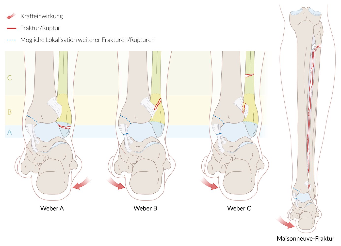
Weber-Klassifikation
- Weber A: unterhalb der intakten Syndesmose (blau)
- Weber B: auf Höhe der Syndesmose mit möglicher Läsion (gelb)
- Weber C: oberhalb der rupturierten Syndesmose (grün)
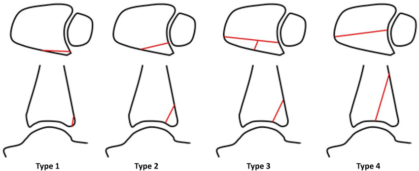
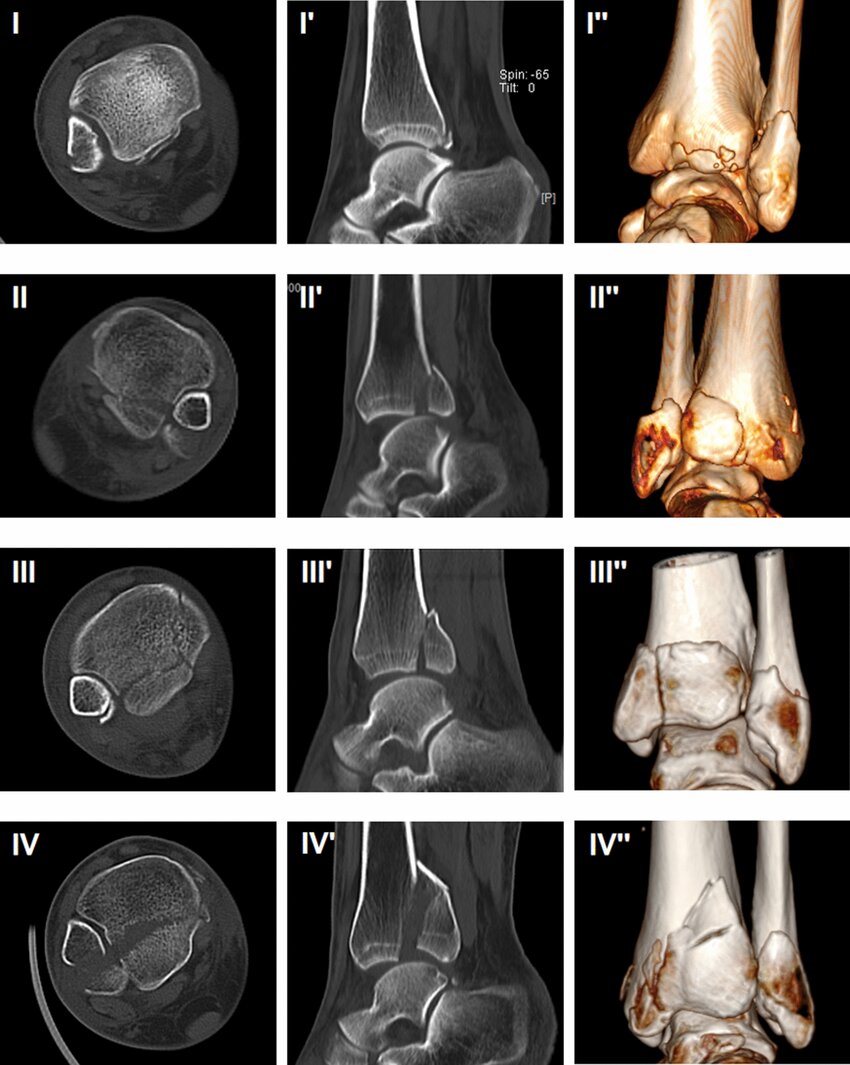
Posteriorer Malleolus
Bartonicek & Rammelt Klassifikation
- Typ 1: Fraktur außerhalb der (intakten) Inzisur (Fibula-Notch)
- Typ 2: Posterolaterales Fragment mit Beteiligung der Inzisur (Fibula-Notch)
- Typ 3: Zweiteiliges, posteromediales Fragment mit Beteiligung des Innenknöchels
- Typ 4: Großes posterolaterales dreieckförmiges Fragment (Beteiligung >⅓ der Inzisur)
- Typ 5: Irregulär, osteoporotisch, keinem der anderen 4 Subtypen zuzuordnen